| Valve and Conduit Replacements |
|
|
 |
|
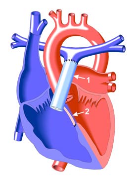
Truncus Arteriosus Repair
Surgical treatment of Truncus Arteriosus involves the separation of the pulmonary arteries from the truncus arteriosus. The pulmonary arteries are then connected to the right ventricle with a valved conduit (light blue in the illustration), usually a pulmonary or aortic homograft (cryopreserved human tissue).
It is important that the patient be monitored for the development of conduit stenosis, or narrowing, which will eventually make reoperation necessary. There are usually no obvious symptoms of this stenosis unless ventricular function becomes impaired, though there may be an increase in fatigue upon exertion and a more intense systolic ejection murmur.
When obstruction is suspected, a catheterization may be performed to determine the degree and location of the narrowing. (Echocardiography is less useful in recognizing conduit obstruction in adults than in children because of imaging problems.)
In addition, the patient should be monitored for the breakdown in function of the pulmonary conduit valve, which may become regurgitant and require replacement.
As the young patient grows, there will be a need for periodic replacements of the conduit. However, after the patient reaches adulthood, the interval between conduit insertion and reoperation is generally between 10 and 20 years. |
|