Hypoplastic Left Heart Syndrome and the Adult Patient
To date, no untreated patients with Hypoplastic Left Heart Syndrome have survived to adulthood. However, dramatic advances in treatment have led to significant improvements in the life expectancy, and treated patients are now surviving into adulthood.
Because of this, general comments about Single Ventricle in the adult are presented below. Many of these concerns and guidelines will apply to adult patients with Hypoplastic Left Heart Syndrome.
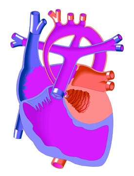
Single Ventricle refers to the congenital heart defects in which the heart functionally has only one pumping chamber. Examples are Tricuspid Atresia, Hypoplastic Left Heart Ventricle, Double Inlet Left Ventricle, and Double Outlet Right Ventricle. Other defects (e.g. some forms of Atrioventricular Canal Defect and Pulmonary Atresia) may create single ventricle conditions in the heart.
Adult patients with these defects will usually have had a Fontan Operation. In cases where early treatment consisted of a Glenn procedure and/or the insertion of a shunt between the systemic circulation and the pulmonary artery (e. g. modified or classic Blalock-Taussig Shunt), the patients may be candidates for the Fontan in later life. Very rarely, a person with Single Ventricle reaches adulthood without treatment and without symptoms. These patients may or may not receive the Fontan, depending on an assessment of the relative risks and benefits.
Most patients who have not had the Fontan Operation will begin to show symptoms of cyanosis (external blueness caused by oxygen-poor arterial blood), fatigue, arrhythmias, and/or exercise intolerance, generally because of insufficient blood flow to the lungs through the pulmonary artery. They will also have a heart murmur because of pulmonary stenosis (narrowing of the outflow tract through which blood flows from the heart to the lungs) and/or because of atrioventricular valve dysfunction (the valve that connects the functioning ventricle with an atrium). If these symptoms are severe, the Fontan Operation will not be performed, but only if certain conditions are met:
· pulmonary artery pressure is acceptably low
· the pulmonary arteries are sufficiently well formed
· there is no pulmonary vascular obstructive disease (PVOD)
· the systemic ventricle is functioning adequately
These criteria will be evaluated through a variety of tests. A chest x-ray, echocardiogram, and MRI (Magnetic Resonance Imaging) will show left ventricular function and other aspects of anatomy and cardiovascular condition. An electrocardiogram is used to check for the presence of arrhythmias (irregular heartbeats) and as an indicator of certain anatomical defects. In addition, a cardiac catheterization procedure will be used to take hemodynamic measurements (blood pressures and concentrations of oxygen and other gases) in the pulmonary arteries and to evaluate their structure.
In some cases where a Fontan operation is possible, the atrioventricular valve will need to be repaired or replaced. The prognosis after this operation for single ventricle patients is better than for other treatments, and improvements of surgical technique continue to be made. Life-long medical monitoring, including the prescription of antibiotics to guard against endocarditis (infection of the heart's internal lining), will be necessary for all single ventricle patients. |